Ultrasounds explained
There seems to be a lot of confusion on types of ultrasounds. People who have suspected a blood clot and visited the Emergency Department often say, ‘I already got an ultrasound!’ So really there are two different types of ultrasounds for veins, each which looks for a different vein disease.
If you go to the Emergency Department because you think you might have a blood clot, the ED is going to perform an ultrasound to rule out Deep Vein Thrombosis. They are looking to see if there is a blood clot or not.
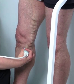
The second kind of ultrasound is for patients who have varicose veins. Varicose veins are due to venous blood flowing the wrong way back down your leg. The flow problem causing varicose veins, however, is often more extensive than we can see just looking at your leg. During a venous insufficiency ultrasound, the ultrasonographer maps the flow in all the major veins in patient’s leg, marking where the blood is flowing in the wrong direction (reflux), noting how the veins connect with this specific patient, where the problem starts and stops, and noting vein diameters and presence or absence of reflux. We use this mapping as a road map to treat the patient, and we also use it to get approval from insurance. If the measurements don’t meet insurance criteria, then insurance does not consider treatment ‘medically necessary’. This venous insufficiency study takes about an hour to perform and is a different study looking for a different disease than the Deep Vein Thrombosis study.
Generally, if a patient has had a venous insufficiency study in the past but it has been over 12 months, then insurance will not accept the findings and will make the patient get a new study. This isn’t a bad idea, since varicose veins are a progressive disease and things often worsen over time. The picture may look worse a year or two later without treatment.